Laparoscopy
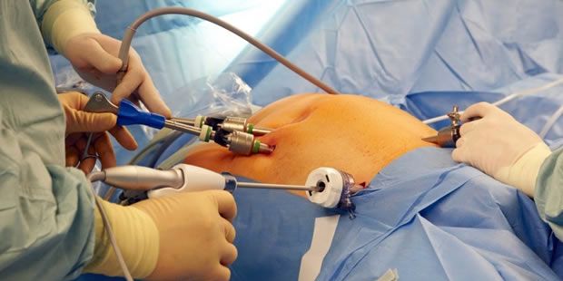
If the patient wishes to become pregnant, but has difficulty conceiving, laparoscopy may allow the physician to identify and sometimes even correct the condition.
Through endoscopic vision, the surgeon can verify the patency of the Fallopian tube (through which the fertilized egg passes).
Particularly, the presence of pelvic endometriosis or adhesions, it cannot be assessed with any other way. Even the status and function of the fimbria, that is, the most distal portion of the tube used to capture ovule, can only be properly highlighted by laparoscopy.
During the laparoscopy in addition to the normal view of all the pelvic organs involved in the reproductive function (uterus, tube, ovary, peritoneum) some procedures are routine while others are carried out only if necessary.
In particular chromosalpingoscopy is always executed, it consists in the injection, through the cervical canal and, therefore, in the vaginal tube, of a vital dye (methylene blue). The filling of the tube and passage through fimbriae in the peritoneum is observed. It is basically a test of tubal patency.
In extreme cases, in women who have already undergone reproductive medicine techniques (IVF / ICSI), where the pathology has irreversibly compromised the tubal functionality, there is an absolute indication to remove one or both tubes (salpingectomy).
Adnexial diseases
Conservative treatment
- Ovarian biopsy
- ovarian drill
- ovarian or falloppian tube cyst removal
- ectopic pregnancy removal
- adhesiolisis
- PID
Demolitive treatment
- ovariectomy
- salpingectomy
- adnexectomy
Endometriosis
It represents one of the best indications to a laparoscopy. Cases of minimal, mild, moderate or severe, endometriosis, even if the endometriosic cysts or nodules are large, can be successfully treated.
Endometriosis is a mysterious disease that affects millions of women, in reproductive age (3-10%), all over the world.
It is rare before menarche and tends to decrease significantly after menopause.
The name derives from the word "endometrium", i.e. the fabric which covers the inner surface of the uterus, and that grows and flakes off every month during the menstrual cycle.
In endometriosis, tissue that is histologically equal to endometrium, is most often located outside the uterus and in other areas of the body.
In such places the endometrial tissue develops in the form of cysts, nodules, lesions, implants or excrescences that are located most frequently in the pelvis, affecting the ovaries, the tube, the uterus ligaments, the area between the vagina and rectum, the outer lining of the uterus and that of the abdominal cavity (peritoneum).
Sometimes these lesions are also found in post-surgical abdominal scars, on the tenuous intestine or rectum-sigma, near ureteri, on the bladder, the vagina, the cervix and vulva.
As the coating of the uterus, endometriosic lesions are usually sensitive to the hormones of the menstrual cycle. Each month they develop, flake, and cause bleeding.
But in these cases the endometrial tissue has no way of escaping outside of the body causing internal bleeding with subsequent alteration of blood and flaked tissue from the injury, inflammation of the surrounding areas and formation of scar tissue.
Other complications can be the breaking of these lesions (which can cause it to spread to other areas), the formation of adhesions, bleeding, intestinal obstruction (for intestinal localization) or urinary problems (for location in the urethra or bladder).
The symptoms that occur more frequently in endometriosis are:
- Pain before and during menstruation (dysmenorrhea)
- Pain during sexual intercourse (dyspareunia)
- Pain during defecation (disketia)
- Diarrhea and / or constipation
- Pain during urination (dysuria)
- Chronic pelvic pain
- Blood in the stool (haematochezia / rectorragy)
- Blood in the urine (hematuria)
- Nausea / vomiting
- Menstrual irregularities
Some women with endometriosis my not show any symptoms. Infertility affects approximately 30-40% of women with endometriosis and is a common outcome with the progression of the disease. The intensity of pain is not related to the size or extent of injuries. Small formations (called petechial) have proven to be more active in the production of prostaglandins (substances that trigger the inflammatory processes) that may explain the significant symptoms that often accompany the presence of small masses. Prostaglandins are substances found in the whole body, involved in numerous functions, and considered responsible for most of the symptoms of endometriosis.
Symptoms often tend to increase over time, although in some cases there are cycles of remission and recurrence.
The staging of the disease must be performed during surgery. There are 4 stages according to the American Fertility Society (rAFS )classification:
Stage I (minimal disease)
Isolated endometriosic tissue, adhering outside the uterine wall, the tube and ovary.
Stage II (mild disease)
There is not always a correlation between the amount of pain and the degree of seriousness of endometriosis. Mild endometriosis may already cause severe pain.
Stage III (moderate disease)
Cysts can form in the ovaries and collect menstrual blood. When an incision is made to these cysts during a surgery, this blood appears as a viscous mass of brownish color. For this reason these cysts are also called chocolate cysts.
Stage IV (severe disease)
The cyclic bleeding of the endometriosic outbreak, stimulate continuous irritation of the peritoneum resulting in the formation of scars (adhesions) and endometriosic nodules that can penetrate the organs and adjacent structures such as bowel, bladder, ureteri, vagina: spreading into the abdominal cavity. In the severe form the rectum-vaginal septum is also involved. In advanced endometrosis, pain is generally stronger. However, it is certainly possible that endometriosis may not result in any significant pain.
Myomectomy
The myoma is a single or multiple benign growth, made of fibrous muscle tissue that can develop on the outer surface or wall of the uterus and can reach the uterine cavity when it is very deep. Its size can sometimes be considerable.
The myomectomy, namely the removal of one or more fibroids (multiple myomectomy) is the more conservative surgery for the treatment for this pathology and aims to restoring the anatomical and functional integrity of the uterus. Myomas or fibroids (similar terms) grow by creating a capsule on their surface that separates them very well from the surrounding tissue and this makes them often easy to remove. The submucosal myoma is the main limiting factor of this technique and is a condition that increases the difficulty of the operation, the risks of complications producing an unsatisfactory surgical result, and so a classification in three degrees has been universally adopted.
Stage 0 myoma develops in the endometrial cavity and has no intramural extension. For this reason, they are easily removed regardless of size and this treatment has the highest success rate and the lowest number of complications.
Intramural myoma develops within the uterine wall but has an extension in the wall which is less than 50% of the volume of myoma defined in stage 1.
Submucosal mayomas have less than half of their volume in the endometrial cavity and having an intramural extension of more than 50% are defined stage 2.
In any case, the thickness of the myometrium between the outer edge of the myoma and the serous surface of the uterus should not be less than 5 mm. This thickness, called free miometrium margin, is measured by ultrasound during the presurgical examination of the patient. The increase in the degree of intramural extension increases the duration of the surgery, technical difficulties, the risk of incomplete removal of the myoma, the risk of intravascularization (passage of the uterus distention liquid into the intravascular compartment of the patient), the risk of uterine perforation and of bleeding.
In some cases prior administration of certain drugs called GnRH analogues may be necessary to reduce the volume and vascularization of the mioma, make it easier and safer to remove.
Hysterectomy
With the laparoscopic hysterectomy the uterus can be completely removed (and the ovaries and tubes if necessary) also if large. The uterine manipulator used to perform the laparoscopic hysterectomy, is different from the traditional one. It is larger and has a series of silicone valves to detach the uterus without losing the gas pressure inside the abdomen that would otherwise escape through the vagina. The uterus, once detached during the laparoscopy, is removed through the vagina and the dome of the vagina is closed with a continuous suture. To the hysterectomy we can also add routine repairs of defects in the pelvic floor (cystocele, urethrocele, rectocele) or surgeries to prevent vaginal vault prolapse. In hysterectomy surgeries for large wombs, a higher incision is needed (a few centimeters above the naval).
Pelvic Adhesions
Often secondary to laparotomic surgery or PID, adhesions are common cause (12%) of infertility and chronic pelvic pain. Sometimes many surgical procedures are needed to solve these pathologies with high social and economic costs. Laparoscopic surgery permits to erase almost all pelvic adhesions with a short hospitalization and a low risk of adesions recurrence.
Endoscopica Malzoni is the only italian center associated to Adhesion related Disorder (ARD) Education and Awareness (www.adhesionrelateddisorder.com), an international society with the porpose of selecting and promoting best adhesion managing centers wordwide.
Pelvic floor defects
Incontinence is a personal and social problem. Involontary urinary loss by coughing is called "stress incontinence". Urge urinary need is called "urge incontinence". While urge incontinence terapy is medical, stress incontinence terapy is surgery. Common surgery was, until 10 years ago, vaginal plastic repair,affected to high recidive odds, replaced today by urethropexy sec.Burch. - Vaginal Sling (T.V.T, T.O.T.) - Laparoscopic Sacro-colpopexy - Laparoscopic Paravaginal repair - Laparoscopic rectocele repair.
Oncologic Pathologies
Currently, laparoscopy is increasingly used for cancer in oncologic centers. Making the pelvic and lombo-aortic linfoadenectomy by endoscopy allows to study and / or treat the initial cases (early stages) of carcinoma of the uterine cervix and the endomyosis, associating it to hysterectomy which is referred to as being "radical". The surgical staging of ovarian cancer (first stage) or the revaluation (second-look) is still possible after cytoreductive surgery in advanced stages. The surgical results that are obtained in terms of radicalism from an oncologic point of view, are generally considered quite consistent with those of traditional surgery.
The patients, in addition to the common benefits of laparoscopy, also get a psychological advantage as they can usually start, very early, any adjuvant treatments (chemo or radiotherapy).
To note, that to date only a small percentage of operations for oncologic diseases in early stages, are executable with the laparoscopic technique.


